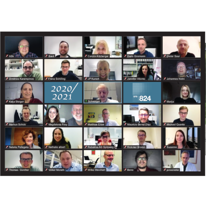
Forschungsbereich B
Mesenchymal stem cells and glioma cells form a structural as well as a functional syncytium in vitro
01.03.2012
Schnell O, Romagna A, Jaehnert I, Albrecht V, Eigenbrod S, Juerchott K, Kretzschmar H, Tonn JC, Schichor C.
Exp Neurol.,
2012,
234(1),
208-19
publiziert am 01.03.2012
The interaction of human mesenchymal stem cells (hMSCs) and tumor cells has been investigated in various contexts. HMSCs are considered as cellular treatment vectors based on their capacity to migrate towards a malignant lesion. However, concerns about unpredictable behavior of transplanted hMSCs are accumulating. In malignant gliomas, the recruitment mechanism is driven by glioma-secreted factors which lead to accumulation of both, tissue specific stem cells as well as bone marrow derived hMSCs within the tumor. The aim of the present work was to study specific cellular interactions between hMSCs and glioma cells in vitro. We show, that glioma cells as well as hMSCs differentially express connexins, and that they interact via gap-junctional coupling. Besides this so-called functional syncytium formation, we also provide evidence of cell fusion events (structural syncytium). These complex cellular interactions led to an enhanced migration and altered proliferation of both, tumor and mesenchymal stem cell types in vitro. The presented work shows that glioma cells display signs of functional as well as structural syncytium formation with hMSCs in vitro. The described cellular phenomena provide new insight into the complexity of interaction patterns between tumor cells and host cells. Based on these findings, further studies are warranted to define the impact of a functional or structural syncytium formation on malignant tumors and cell based therapies in vivo.
Copyright © 2011 Elsevier Inc. All rights reserved.
Cryptate mediated nucleophilic 18F-fluorination without azeotropic drying
16.02.2012
Wessmann SH, Henriksen G, Wester HJ.
Nuklearmedizin.,
2012,
50(1),
1-8
publiziert am 16.02.2012
The radiosynthesis of the vast majority of 18F-labeled tracers rely on azeotropic drying of [18F]fluoride and subsequent cryptate mediated introduction of [18F]fluoride by nucleophilic substitution. The aim of this study was to develop a method for simplification of this process, based on preparation of reactive [K+⊂2.2.2]18F- by solvent drying of [18F]fluoride adsorbed onto an anion exchange resin. Methods: Aqueous [18F]fluoride (0.5-1 ml) obtained from the 18O(p,n)18F nuclear reaction was trapped on a strong anion-exchange (SAX) cartridge. After washing the cartridge with dry CH3CN, [18F]fluoride was eluted with an anhydrous solution of [K+⊂2.2.2]OH- in CH3CN and directly used for nucleophilic fluorination reactions. Results: [18F]Fluoride from target water was quantitatively retained by the SAX cartridge, and water-free [18F]fluoride recovered in an overall yield of 92±5% (n = 10). [18F]Fluoride obtained by this procedure led to radiochemical yields of 70-90% for [18F]FDG, [18F]FET, [18F]FLT, [18F]FAZA and [18F]Fallypride. Conclusion: SAX-resin adsorbed [18F]fluoride can be dried with non-aqueous solvents and eluted with [K+⊂2.2.2]OH- in CH3CN. The reactivity of [K+⊂2.2.2]F- generated by the new method is comparable to that of [18F]fluoride obtained by azeotropic drying. The described procedure facilitates the automated production of 18F-radiopharmaceuticals in general, and may also simplify the use of microfluidic devices for 18F-radiotracer production.
Basal HIF-1α expression levels are not predictive for radiosensitivity of human cancer cell lines
12.02.2012
Schilling D, Bayer C, Emmerich K, Molls M, Vaupel P, Huber RM, Multhoff G.
Strahlenther Onkol.,
2012,
publiziert am 12.02.2012
BACKGROUND AND PURPOSE:
High levels of hypoxia inducible factor (HIF)-1α in tumors are reported to be associated with tumor progression and resistance to therapy. To examine the impact of HIF-1α on radioresistance under normoxia, the sensitivity towards irradiation was measured in human tumor cell lines that differ significantly in their basal HIF-1α levels.
MATERIAL AND METHODS:
HIF-1α levels were quantified in lysates of H1339, EPLC-272H, A549, SAS, XF354, FaDu, BHY, and CX- tumor cell lines by ELISA. Protein levels of HIF-1α, HIF-2α, carbonic anhydrase IX (CA IX), and GAPDH were assessed by Western blot analysis. Knock-down experiments were performed using HIF-1α siRNA. Clonogenic survival after irradiation was determined by the colony forming assay.
RESULTS:
According to their basal HIF-1α status, the tumor cell lines were divided into low (SAS, XF354, FaDu, A549, CX-), intermediate (EPLC-272H, BHY), and high (H1339) HIF-1α expressors. The functionality of the high basal HIF-1α expression in H1339 cells was proven by reduced CA IX expression after knocking-down HIF-1α. Linear regression analysis revealed no correlation between basal HIF-1α levels and the survival fraction at either 2 or 4 Gy in all tumor cell lines investigated.
CONCLUSION:
Our data suggest that basal HIF-1α levels in human tumor cell lines do not predict their radiosensitivity under normoxia.
Krüppel-like factor 8 (KLF8) is expressed in gliomas of different WHO grades and is essential for tumor cell proliferation
19.01.2012
Schnell O, Romagna A, Jaehnert I, Albrecht V, Eigenbrod S, Juerchott K, Kretzschmar H, Tonn JC, Schichor C.
PLoS One.,
2012,
7(1),
e30429
publiziert am 19.01.2012
Krüppel-like factor 8 (KLF8) has only recently been identified to be involved in tumor cell proliferation and invasion of several different tumor entities like renal cell carcinoma, hepatocellular carcinoma and breast cancer. In the present study, we show for the first time the expression of KLF8 in gliomas of different WHO grades and its functional impact on glioma cell proliferation. In order to get information about KLF8-mRNA regulation qPCR was performed and did not reveal any significant difference in samples (n = 10 each) of non-neoplastic brain (NNB), low-grade gliomas (LGG, WHO°II) and glioblastomas (GBM, WHO°IV). Immunohistochemistry of tissue samples (n = 7 LGG, 11 AA and 12 GBM) did not show any significant difference in the fraction of KLF8-immunopositive cells of all analyzed cells in LGG (87%), AA (80%) or GBM (89%). Tissue samples from cerebral breast cancer metastasis, meningiomas but also non-neoplastic brain demonstrated comparable relative cell counts as well. Moreover, there was no correlation between KLF8 expression and the expression pattern of the assumed proliferation marker Ki67, which showed high variability between different tumor grade (9% (LGG), 6% (AA) and 15% (GBM) of Ki67-immunopositive cells). Densitometric analysis of Western blotting revealed that the relative amount of KLF8-protein did also not differ between the highly aggressive and proliferative GBM (1.05) compared to LGG (0.93; p<0.05, studens t-test). As demonstrated for some other non-glial cancer entities, KLF8-knockdown by shRNA in U87-MG cells confirmed its functional relevance, leading to an almost complete loss of tumor cell proliferation. Selective blocking of KLF8 might represent a novel anti-proliferative treatment strategy for malignant gliomas. Yet, its simultaneous expression in non-proliferating tissues could hamper this approach.
Chronic inflammation in cancer development
12.01.2012
Gabriele Multhoff, Michael Molls and Jürgen Radons
frontiers in immunology,
2012,
doi: 10.3389,
fimmu.201100098
publiziert am 12.01.2012
Chronic inflammatory mediators exert pleiotropic effects in the development of cancer. On the one hand, inflammation favors carcinogenesis, malignant transformation, tumor growth, invasion, and metastatic spread; on the other hand inflammation can stimulate immune effector mechanisms that might limit tumor growth. The linkbetween cancer and inflammation depends on intrinsic and extrinsic pathways. Both pathways result in the activation of transcription factors such as NF-KB, STAT-3, and HIF-1 and in accumulation of tumorigenic factors in tumor and microenvironment. STAT-3 and NF-KB interact at multiple levels and thereby boost tumor-associated inflammation which can suppress anti-tumor immune responses. These factors also promote tumor growth, progression, and metastatic spread. IL-1, IL-6, TNF, and PGHS-2 are key mediators of an inflammatory milieu by modulating the expression of tumor-promoting factors. In this review we concentrate on the crucial role of pro-inflammatory mediators in inflammation-driven carcinogenesis and outline molecular mechanisms of IL-1 signaling in tumors. In addition, we elucidate the dual roles of stress proteins äs danger Signals in the development of anti-cancer immunity and anti-apoptotic functions.
TFAP2E–DKK4 and Chemoresistance in Colorectal Cancer
05.01.2012
Ebert MP, Tänzer M, Balluff B, Burgermeister E, Kretzschmar AK, Hughes DJ, Tetzner R, Lofton-Day C, Rosenberg R, Reinacher-Schick AC, Schulmann K, Tannapfel A, Hofheinz R, Röcken C, Keller G, Langer R, Specht K, Porschen R, Stöhlmacher-Williams J, Schuster T, Ströbel P, Schmid RM
N Engl J Med.,
2012,
366(1),
44-53
publiziert am 05.01.2012
Background:
Chemotherapy for advanced colorectal cancer leads to improved survival; however, predictors of response to systemic treatment are not available. Genomic and epigenetic alterations of the gene encoding transcription factor AP-2 epsilon (TFAP2E) are common in human cancers. The gene encoding dickkopf homolog 4 protein (DKK4) is a potential downstream target of TFAP2E and has been implicated in chemotherapy resistance. We aimed to further evaluate the role of TFAP2E and DKK4 as predictors of the response of colorectal cancer to chemotherapy.
Methods:
We analyzed the expression, methylation, and function of TFAP2E in colorectal-cancer cell lines in vitro and in patients with colorectal cancer. We examined an initial cohort of 74 patients, followed by four cohorts of patients (total, 220) undergoing chemotherapy or chemoradiation.
Results:
TFAP2E was hypermethylated in 38 of 74 patients (51%) in the initial cohort. Hypermethylation was associated with decreased expression of TFAP2E in primary and metastatic colorectal-cancer specimens and cell lines. Colorectal-cancer cell lines overexpressing DKK4 showed increased chemoresistance to fluorouracil but not irinotecan or oxaliplatin. In the four other patient cohorts, TFAP2E hypermethylation was significantly associated with nonresponse to chemotherapy (P<0.001). Conversely, the probability of response among patients with hypomethylation was approximately six times that in the entire population (overall estimated risk ratio, 5.74; 95% confidence interval, 3.36 to 9.79). Epigenetic alterations of TFAP2E were independent of mutations in key regulatory cancer genes, microsatellite instability, and other genes that affect fluorouracil metabolism.
Conclusions:
TFAP2E hypermethylation is associated with clinical nonresponsiveness to chemotherapy in colorectal cancer. Functional assays confirm that TFAP2E-dependent resistance is mediated through DKK4. In patients who have colorectal cancer with TFAP2E hypermethylation, targeting of DKK4 may be an option to overcome TFAP2E-mediated drug resistance. (Funded by Deutsche Forschungsgemeinschaft and others.).
PET of CXCR4 expression by a (68)Ga-labeled highly specific targeted contrast agent.
01.11.2011
Gourni E, Demmer O, Schottelius M, D'Alessandria C, Schulz S, Dijkgraaf I, Schumacher U, Schwaiger M, Kessler H, Wester HJ.
J Nucl Med.,
2011,
52(11),
1803-10
publiziert am 01.11.2011
The overexpression of the chemokine receptor CXCR4 plays an important role in oncology, since together with its endogenous ligand, the stromal cell-derived factor (SDF1-α), CXCR4 is involved in tumor development, growth, and organ-specific metastasis. As part of our ongoing efforts to develop highly specific CXCR4-targeted imaging probes and with the aim to assess the suitability of this ligand for first proof-of-concept studies in humans, we further evaluated the new (68)Ga-labeled high-affinity cyclic CXCR4 ligand, (68)Ga-CPCR4-2 (cyclo(D-Tyr(1)-[NMe]-D-Orn(2)-[4-(aminomethyl) benzoic acid,(68)Ga-DOTA]-Arg(3)-2-Nal(4)-Gly(5))).
METHODS:
Additional biodistribution and competitions studies in vivo, dynamic PET studies, and investigations on the metabolic stability and plasma protein binding were performed in nude mice bearing metastasizing OH1 human small cell lung cancer xenografts. CXCR4 expression on OH1 tumor sections was determined by immunohistochemical staining.
RESULTS:
(nat)Ga-CPCR4-2 exhibits high CXCR4 affinity with a half maximum inhibitory concentration of 4.99 ± 0.72 nM. (68)Ga-CPCR4-2 showed high in vivo stability and high and specific tumor accumulation, which was reduced by approximately 80% in competition studies with AMD3100. High CXCR4 expression in tumors was confirmed by immunohistochemical staining. (68)Ga-CPCR4-2 showed low uptake in nontumor tissue and particularly low kidney accumulation despite predominant renal excretion, leading to high-contrast delineation of tumors in small-animal PET studies.
CONCLUSION:
The small and optimized cyclic peptide CPCR4-2 labeled with (68)Ga is a suitable tracer for targeting and imaging of human CXCR4 receptor expression in vivo. The high affinity for CXCR4, its in vivo stability, and the excellent pharmacokinetics recommend the further evaluation of (68)Ga-CPCR4-2 in a proof-of-concept study in humans.
MALDI imaging identifies prognostic seven-protein signature of novel tissue markers in intestinal-type gastric cancer
18.10.2011
Balluff B, Rauser S, Meding S, Elsner M, Schöne C, Feuchtinger A, Schuhmacher C, Novotny A, Jütting U, Maccarrone G, Sarioglu H, Ueffing M, Braselmann H, Zitzelsberger H, Schmid RM, Höfler H, Ebert MP, Walch A.
Am J Pathol.,
2011,
179(6),
2720-9
publiziert am 18.10.2011
Proteomics-based approaches allow us to investigate the biology of cancer beyond genomic initiatives. We used histology-based matrix-assisted laser desorption/ionization (MALDI) imaging mass spectrometry to identify proteins that predict disease outcome in gastric cancer after surgical resection. A total of 181 intestinal-type primary resected gastric cancer tissues from two independent patient cohorts were analyzed. Protein profiles of the discovery cohort (n = 63) were directly obtained from tumor tissue sections by MALDI imaging. A seven-protein signature was associated with an unfavorable overall survival independent of major clinical covariates. The prognostic significance of three individual proteins identified (CRIP1, HNP-1, and S100-A6) was validated immunohistochemically on tissue microarrays of an independent validation cohort (n = 118). Whereas HNP-1 and S100-A6 were found to further subdivide early-stage (Union Internationale Contre le Cancer [UICC]-I) and late-stage (UICC II and III) cancer patients into different prognostic groups, CRIP1, a protein previously unknown in gastric cancer, was confirmed as a novel and independent prognostic factor for all patients in the validation cohort. The protein pattern described here serves as a new independent indicator of patient survival complementing the previously known clinical parameters in terms of prognostic relevance. These results show that this tissue-based proteomic approach may provide clinically relevant information that might be beneficial in improving risk stratification for gastric cancer patients.
Copyright © 2011 American Society for Investigative Pathology. Published by Elsevier Inc. All rights reserved.
A novel expression and purification system for the production of enzymatic and biologically active human granzyme B
31.08.2011
Stangl S, Themelis G, Friedrich L, Ntziachristos V, Sarantopoulos A, Molls M, Skerra A, Multhoff G.
J Immunol Methods.,
2011,
371(1-2),
8-17
publiziert am 31.08.2011
The serine protease granzyme B (grB) has previously been shown to induce perforin-independent apoptosis in membrane Hsp70 positive tumor cells in a number of in vitro experimental systems. Ongoing studies that are investigating the in vivo relevance of these findings by assessing the therapeutic potential of grB in a human xenograft tumor mouse model required the development of an expression system for the production of high yields of enzymatic and biologically active human grB. In order to maintain potentially important posttranslational modifications that occur in mammalian cells, human embryonal kidney cells (HEK293) were stably transfected with human grB. The HEK293 host cells were protected from apoptotic cell death by fusing an inactivation site coupled to a (His)(6) tag to the gene sequence of GrB. Inactive grB which was actively released from HEK293 cells by insertion of a Igκ leader sequence was purified on a nickel column utilizing the (His)(6) tag. After enterokinase digestion and heparin affinity chromatography, high yields of enzymatic and biologically active human grB were obtained. The perforin-independent interaction of grB with membrane Hsp70 positive tumor cells appeared to be associated with mammalian glycosylation and mediated by the oligosaccharide moiety of neuraminic acid (NANA).
Copyright © 2011. Published by Elsevier B.V.
(18)F-FDG PET-guided salvage neoadjuvant radiochemotherapy of adenocarcinoma of the esophagogastric junction: the MUNICON II trial
01.08.2011
zum Büschenfelde CM, Herrmann K, Schuster T, Geinitz H, Langer R, Becker K, Ott K, Ebert M, Zimmermann F, Friess H, Schwaiger M, Peschel C, Lordick F, Krause BJ.
J Nucl Med.,
2011,
52(8),
1189-96
publiziert am 01.08.2011
Previous studies demonstrated that chemotherapy-induced changes in tumor glucose metabolism measured with (18)F-FDG PET identify patients who benefit from preoperative chemotherapy and those who do not. The prognosis for chemotherapy metabolic nonresponders is poorer than for metabolic responders. Therefore, we initiated this prospective trial to improve the clinical outcome of metabolic nonresponders using a salvage neoadjuvant radiochemotherapy.
METHODS:
Fifty-six patients with locally advanced adenocarcinomas of the esophagogastric junction were included. Tumor glucose uptake was assessed by (18)F-FDG PET before chemotherapy and 14 d after initiation of chemotherapy. PET nonresponders received salvage neoadjuvant radiochemotherapy, whereas metabolic responders received neoadjuvant chemotherapy for 3 mo before surgery.
RESULTS:
Thirty-three patients were metabolic responders, and 23 were nonresponders. Resection was performed on 54 patients. R0 resection rate was 82% (95% confidence interval [CI], 66%-91%) in metabolic responders and 70% (95% CI, 49%-84%) in metabolic nonresponders (P = 0.51). Major histologic remissions were observed in 12 metabolic responders (36%; 95% CI, 22%-53%) and 6 nonresponders (26%; 95% CI, 13%-46%). One-year progression-free rate was 74% ± 8% in PET responders and 57% ± 10% in metabolic nonresponders (log rank test, P = 0.035). One-year overall survival was comparable between the groups (∼80%), and 2-y overall survival was estimated to be 71% ± 8% in metabolic responders and 42% ± 11% in PET nonresponders (hazard ratio, 1.9; 95% CI, 0.87-4.24; P = 0.10).
CONCLUSION:
This prospective study showed the feasibility of a PET-guided treatment algorithm. However, by comparing the groups of nonresponding patients in the current trial and the previous published MUNICON (Metabolic response evalUatioN for Individualisation of neoadjuvant Chemotherapy in Esophageal and esophagogastric adeNocarcinoma) I trial, increased histopathologic response was observed after salvage radiochemotherapy, but the primary endpoint of the study to increase the R0 resection rate was not met. The prognosis of the subgroup of PET nonresponders remains poor, indicating their different tumor biology.
Bevacizumab can induce reactivity to VEGF-C and -D in human brain and tumour derived endothelial cells
01.08.2011
Grau S, Thorsteinsdottir J, von Baumgarten L, Winkler F, Tonn JC, Schichor C.
J Neurooncol.,
2011,
104(1),
103-12
publiziert am 01.08.2011
Though clinical trials demonstrated effectiveness of the anti-VEGF antibody bevacizumab (Avastin) in adjuvant therapies for some solid tumours, there are rather few experimental data about cellular effects of bevacizumab on tumour cells and tumour associated endothelial cells. Recent reports demonstrate resistance mechanisms and secondary re-angiogenesis after a transient normalization of tumour vessels. Therefore we investigated the influence of bevacizumab on human glioma cells and human brain derived as well as tumour derived endothelial cells focussing on the role of VEGF-C and -D as potential alternative pro-angiogenic factors. Bevacizumab treatment showed no influence on proliferation after short term exposure (1-5 days) but slowed down endothelial cell proliferation by 25-30% after 14 days treatment. There was no significant induction of apoptosis after short or long term exposure. Tube formation capabilities were significantly impaired by bevacizumab with a continuing effect after 14 days of treatment even after omitting the antibody. VEGF-C and -D had no effect on endothelial cells in untreated or short term treatment groups. However, cells developed responsiveness to these factors in terms of increased proliferation and tube formation after 14 days bevacizumab treatment. Furthermore, bevacizumab induced expression of VEGF-C and -D in glioma cells. Treatment with bevacizumab may induce alterations in human brain and tumour endothelial cells leading to escape mechanisms from anti-VEGF therapy. VEGF-C and -D thus might act as alternative pro-angiogenic factors during anti-VEGF therapy.
Detection of irradiation-induced, membrane heat shock protein 70 (Hsp70) in mouse tumors using Hsp70 Fab fragment
23.06.2011
Stangl S, Themelis G, Friedrich L, Ntziachristos V, Sarantopoulos A, Molls M, Skerra A, Multhoff G.
Radiother Oncol.,
2011,
99(3),
313-6
publiziert am 23.06.2011
BACKGROUND AND PURPOSE:
The major stress-inducible heat shock protein 70 (Hsp70) is frequently overexpressed in highly aggressive tumors, and elevated intracellular Hsp70 levels mediate protection against apoptosis. Following therapeutic intervention, such as ionizing irradiation, translocation of cytosolic Hsp70 to the plasma membrane is selectively increased in tumor cells and therefore, membrane Hsp70 might serve as a therapy-inducible, tumor-specific target structure.
MATERIALS AND METHODS:
Based on the IgG1 mouse monoclonal antibody (mAb) cmHsp70.1, we produced the Hsp70-specific recombinant Fab fragment (Hsp70 Fab), as an imaging tool for the detection of membrane Hsp70 positive tumor cells in vitro and in vivo.
RESULTS:
The binding characteristics of Hsp70 Fab towards mouse colon (CT26) and pancreatic (1048) carcinoma cells at 4 °C were comparable to that of cmHsp70.1 mAb, as determined by flow cytometry. Following a temperature shift to 37 °C, Hsp70 Fab rapidly translocates into subcellular vesicles of mouse tumor cells. Furthermore, in tumor-bearing mice Cy5.5-conjugated Hsp70 Fab, but not unrelated IN-1 control Fab fragment (IN-1 ctrl Fab), gradually accumulates in CT26 tumors between 12 and 55 h after i.v. injection.
CONCLUSIONS:
In summary, the Hsp70 Fab provides an innovative, low immunogenic tool for imaging of membrane Hsp70 positive tumors, in vivo.
Copyright © 2011 Elsevier Ireland Ltd. All rights reserved.
Targeting membrane heat-shock protein 70 (Hsp70) on tumors by cmHsp70.1 antibody
11.01.2011
Stefan Stangl, Mathias Gehrmann, Julia Riegger, Kristin Kuhs, Isabelle Riederer, Wolfgang Sievert, Kathrin Hubea, Ralph Mocikat, Ralf Dressel, Elisabeth Kremmer, Alan G. Pockleyd, Lars Friedrich, Laszlo Vigh, Arne Skerra and Gabriele Multhoff
Proc Natl Acad Sci U S A,
2011,
108(2),
733-8
publiziert am 11.01.2011
Immunization of mice with a 14-mer peptide TKDNNLLGRFELSG, termed “TKD,” comprising amino acids 450–461 (aa450–461) in the C terminus of inducible Hsp70, resulted in the generation of an IgG1 mouse mAb cmHsp70.1. The epitope recognized by cmHsp70.1mAb, which has been confirmed to be located in the TKD sequence by SPOT analysis, is frequently detectable on the cell surface of human and mouse tumors, but not on isogenic cells and normal tissues, and membrane Hsp70 might thus serve as a tumor-specific target structure. As shown for human tumors, Hsp70 is associated with cholesterol-rich microdomains in the plasma membrane of mouse tumors.
Herein, we show that the cmHsp70.1 mAb can selectively induce antibody-dependent cellular cytotoxicity (ADCC) of membrane Hsp70+ mouse tumor cells by unstimulated mouse spleen cells. Tumor killing could be further enhanced by activating the effector cells with TKD and IL-2. Three consecutive injections of the cmHsp70.1 mAb into mice bearing CT26 tumors significantly inhibited tumor growth and enhanced the overall survival. These effects were associated with infiltrations of NK cells, macrophages, and granulocytes. The Hsp70 specificity of the ADCC response was confirmed by preventing the antitumor response in tumor-bearing mice by coinjecting the cognate TKD peptide with the cmHsp70.1 mAb, and by blocking the binding of cmHsp70.1 mAb to CT26 tumor cells using either TKD peptide or the C-terminal substrate-binding domain of Hsp70.
Distinguishing integral and receptor-bound heat shock protein 70 (Hsp70) on the cell surface by Hsp70 specific antibodies
17.12.2010
Gabriele Multhoff, Lawrence E. Hightower
Cell Stress Chaperones.,
2010,
publiziert am 17.12.2010
Cell Stress & Chaperones journal has become a major outlet for papers and review articles about anti-heat shock protein (HSP) antibodies. In the last decade it became evident that apart from their intracellular localization, members of the heat shock protein 90 (HSP90, HSPC) and HSP70 (HSPA) family are also found on the cell surface. In this review we will focus on Hsp70 (HSPA1A) the major stress-inducible member of the human HSP70 family. Depending on the cell type, the membrane association of Hsp70 comes in two forms. In tumor cells Hsp70 appears to be integrated within the plasma membrane, whereas in non-malignantly transformed (herein termed normal) cells Hsp70 is associated with cell surface receptors. This observation raises the question whether or not these two surface forms of Hsp70 in tumor and normal cells can be distinguished using Hsp70 specific antibodies. Presently a number of Hsp70 specific antibodies are commercially available. These antibodies were generated by immunizing mice either with recombinant or HeLa-derived human Hsp70 protein, parts of the Hsp70 protein or with synthetic peptides. This review aims to characterize the binding of different anti-human Hsp70 antibodies and their capacity to distinguish between integrated and receptor-bound Hsp70 in tumor and normal cells.
Irradiation-Induced Up-Regulation of HLA-E on Macrovascular Endothelial Cells Confers Protection against Killing by Activated Natural Killer Cells
16.12.2010
Isabelle Riederer, Wolfgang Sievert, Günther Eissner, Michael Molls, Gabriele Multhoff
PLoS One.,
2010,
5,
(12):e15339
publiziert am 16.12.2010
Background
Apart from the platelet/endothelial cell adhesion molecule 1 (PECAM-1, CD31), endoglin (CD105) and a positive factor VIII-related antigen staining, human primary and immortalized macro- and microvascular endothelial cells (ECs) differ in their cell surface expression of activating and inhibitory ligands for natural killer (NK) cells. Here we comparatively study the effects of irradiation on the phenotype of ECs and their interaction with resting and activated NKcells.
Methodology/Principal Findings
Primary macrovascular human umbilical vein endothelial cells (HUVECs) only express UL16 binding protein 2 (ULBP2) and the major histocompatibility complex (MHC) class I chain-related protein MIC-A (MIC-A) as activating signals for NK cells, whereas the corresponding immortalized EA.hy926 EC cell line additionally present ULBP3, membrane heat shock protein 70 (Hsp70), intercellular adhesion molecule ICAM-1 (CD54) and HLA-E. Apart from MIC-B, the immortalized human microvascular endothelial cell line HMEC, resembles the phenotype of EA.hy926. Surprisingly, primary HUVECs are more sensitive to Hsp70 peptide (TKD) plus IL-2 (TKD/IL-2)-activated NK cells than their immortalized ECcounterparts. This finding is most likely due to the absence of the inhibitory ligand HLA-E, since the activating ligands are shared among the ECs. The co-culture of HUVECs with activated NK cells induces ICAM-1 (CD54) and HLA-E expression on the former which drops to the initial low levels (below 5%) when NK cells are removed. Sublethal irradiation of HUVECs induces similar but less pronounced effects on HUVECs. Along with these findings, irradiation also induces HLA-E expression on macrovascular ECs and this correlates with an increased resistance to killing by activated NK cells. Irradiation had no effect on HLA-E expression on microvascular ECs and the sensitivity of these cells to NK cells remained unaffected.
Conclusion/Significance
These data emphasize that an irradiation-induced, transient up-regulation of HLA-E on macrovascular ECs might confer protection against NK cell-mediated vascular injury.
TNF-a respecifies human mesenchymal stem cells to a neural fate and promotes migration toward experimental glioma
03.12.2010
V Egea, L von Baumgarten, C Schichor, B Berninger, T Popp, P Neth, R Goldbrunner, Y Kienast, F Winkler, M Jochum and C Ries
Cell Death and Differentiation advance online publication,
2010,
publiziert am 03.12.2010
Bone marrow-derived human mesenchymal stem cells (hMSCs) have become valuable candidates for cell-based therapeutical applications including neuroregenerative and anti-tumor strategies. Yet, the molecular mechanisms that control hMSC trans-differentiation to neural cells and hMSC tropism toward glioma remain unclear. Here, we demonstrate that hMSCs incubated with 50 ng/ml tumor necrosis factor alpha (TNF-a) acquired astroglial cell morphology without affecting proliferation, which was increased at 5 ng/ml. TNF-a (50 ng/ml) upregulated expression of numerous genes important for neural cell growth and function including LIF (leukemia inhibitory factor), BMP2 (bone morphogenetic protein 2), SOX2 (SRY box 2), and GFAP (glial fibrillary acidic protein), whereas NES (human nestin) transcription ceased suggesting a premature neural phenotype in TNF-a-differentiated hMSCs. Studies on intracellular mitogen-activated protein kinase (MAPK) signaling revealed that inhibition of extracellular signal-regulated kinase 1/2 (ERK1/2) activity abolished the TNF-a-mediated regulation of neural genes in hMSCs. In addition, TNF-a significantly enhanced expression of the chemokine receptor CXCR4 (CXC motive chemokine receptor 4), which facilitated the chemotactic invasiveness of hMSCs toward stromal cell-derived factor 1 (SDF-1) alpha. TNF-a-pretreated hMSCs not only exhibited an increased ability to infiltrate glioma cell spheroids dependent on matrix metalloproteinase activity in vitro, but they also showed a potentiated tropism toward intracranial malignant gliomas in an in vivo mouse model. Taken together, our results provide evidence that culture-expansion of hMSCs in the presence of TNF-a triggers neural gene expression and functional capacities, which could improve the use of hMSCs in the treatment of neurological disorders including malignant gliomas.
Cell Death and Differentiation advance online publication, 3 December 2010; doi:10.1038/cdd.2010.154
Aldehyde dehydrogenase 1 positive glioblastoma cells show brain tumor stem cell capacity
13.07.2010
Michael Rasper, Andrea Schäfer, Guido Piontek, Julian Teufel, Gero Brockhoff, Florian Ringel, Stefan Heindl, Claus Zimmer and Jürgen Schlegel
Neuro Oncol.,
2010,
publiziert am 13.07.2010
Glioblastoma (GBM) is the most aggressive primary brain tumor and is resistant to all therapeutic regimens. Relapse occurs regularly and might be caused by a poorly characterized tumor stem cell (TSC) subpopulation escaping therapy. We suggest aldehyde dehydrogenase 1 (ALDH1) as a novel stem cell marker in human GBM. Using the neurosphere formation assay as a functional method to identify brain TSCs, we show that high protein levels of ALDH1 facilitate neurosphere formation in established GBM cell lines. Even single ALDH1 positive cells give rise to colonies and neurospheres. Consequently, the inhibition of ALDH1 in vitro decreases both the number of neurospheres and their size. Cell lines without expression of ALDH1 do not form tumor spheroids under the same culturing conditions. High levels of ALDH1 seem to keep tumor cells in an undifferentiated, stem cell-like state indicated by the low expression of beta-III-tubulin. In contrast, ALDH1 inhibition induces premature cellular differentiation and reduces clonogenic capacity. Primary cell cultures obtained from fresh tumor samples approve the established GBM cell line results.
Classifiation of HER2/neu Status in Gastric Cancer Using a Breast-Cancer Derived Proteome Classfier
08.06.2010
Benjamin Balluff, Mareike Elsner, Andreas Kowarsch, Sandra Rauser, Stephan Meding, Christoph Schuhmacher, Marcus Feith, Ken Herrmann, Christoph Röcken, Roland M. Schmid, Heinz Höfler, Axel Walch and Matthias P. Ebert
Journal of Proteome Research,
2010,
publiziert am 08.06.2010
HER2-testing in breast and gastric cancers is mandatory for the treatment with trastuzumab. We hypothesized that imaging mass spectrometry (IMS) of breast cancers may be useful for generating a classifier that may determine HER2-status in other cancer entities irrespective of primary tumor site. A total of 107 breast (n 48) and gastric (n 59) cryo tissue samples was analyzed by IMS (HER2 was present in 29 cases). The obtained proteomic profiles were used to create HER2 prediction models using different classification algorithms. A breast cancer proteome derived classifier, with HER2 present in 15 cases, correctly predicted HER2-status in gastric cancers with a sensitivity of 65% and a specificity of 92%. To create a universal classifier for HER2-status, breast and nonbreast cancer samples were combined, which increased sensitivity to 78%, and specificity was 88%. Our proof of principle study provides evidence that HER2-status can be identified on a proteomic level across different cancer types suggesting that HER2 overexpression may constitute a unique molecular event independent of the tumor site. Furthermore, these results indicate that IMS may be useful for the determination of potential drugable targets, as it offers a quicker, cheaper, and more objective analysis than the standard HER2-testing procedures immunohistochemistry and fluorescence in situ hybridization.
In vivo imaging of CT26 mouse tumors by using cmHsp70.1 monoclonal antibody
15.03.2010
Stefan Stangl, Mathias Gehrmann, Ralf Dressel, Frauke Alves, Christian Dullin, George Themelis, Vasilis Ntziachristos, Eva Staeblein, Axel Walch, Isabel Winkelmann, Gabriele Multhoff
Journal of Cellular and Molecular Medicine - Decision on Manuscript ID JCMM,
2010,
publiziert am 15.03.2010
The major stress-inducible heat shock protein 70 (Hsp70) is frequently present on the cell surface of human tumors, but not on normal cells. Herein, the binding characteristics of the cmHsp70.1 mouse monoclonal antibody (mAb) were evaluated in vitro and in a syngeneic tumor mouse model. More than 50% of the CT26 mouse colon carcinoma cells express Hsp70 on their cell surface at 4°C. After a temperature shift to 37°C, the cmHsp70.1-FITC mAb translocates into early endosomes and lysosomes Intraoperative and near-infrared fluorescence (NIRF) imaging revealed an enrichment of Cy5.5-conjugated mAb cmHsp70.1, but not an identically labelled IgG1 isotype-matched control, in i.p. and s.c. located CT26 tumors, as soon as 30min after i.v. injection into the tail vein. Due to the rapid turnover rate of membrane-bound Hsp70, the fluorescence-labelled cmHsp70.1 mAb became endocytosed and accumulated in the tumor, reaching a maximum after 24h and remained detectable at least up to 96h after a single i.v. injection. The tumor-selective internalization of mAb cmHsp70.1 at the physiological temperature of 37°C might enable a targeted uptake of toxins or radionuclides into Hsp70 membrane-positive tumors. The anti-tumoral activity of the cmHsp70.1 mAb is further supported by its capacity to mediate antibody dependent cytotoxicity (ADCC).
Performance of Epigenetic Markers SEPT9 and ALX4 in Plasma for Detection of Colorectal Precancerous Lesions
05.02.2010
Marc Tänzer, Benjamin Balluff, Jürgen Distler, Kari Hale, Andreas Leodolter, Christoph Röcken, Bela Molnar, Roland Schmid, Catherine Lofton-Day, Tibor Schuster, Matthias P. A. Ebert
PLoS One,
2010,
5,
2
publiziert am 05.02.2010
Background: Screening for colorectal cancer (CRC) has shown to reduce cancer-related mortality, however, acceptance and compliance to current programmes are poor. Developing new, more acceptable non-invasive tests for the detection of cancerous and precancerous colorectal lesions would not only allow preselection of individuals for colonoscopy, but may also prevent cancer by removal of precancerous lesions.
Methods: Plasma from 128 individuals (cohort I – exploratory study: 73 cases / 55 controls ) was used to test the performance of a single marker, SEPT9, using a real-time quantitative PCR assay. To validate performance of SEPT9, plasma of 76 individuals (cohort II – validation study: 54 cases / 22 controls) was assessed. Additionally, improvement of predictive capability considering SEPT9 and additionally ALX4 methylation was investigated within these patients.
Results: In both cohorts combined, methylation of SEPT9 was observed in 9% of controls (3/33), 29% of patients with colorectal precancerous lesions (27/94) and 73% of colorectal cancer patients (24/33). The presence of both SEPT9 and ALX4 markers was analysed in cohort II and was observed in 5% of controls (1/22) and 37% of patients with polyps (18/49). Interestingly, also 3/5 (60%) patients with colorectal cancer were tested positive by the two marker panel in plasma.
Conclusions: While these data confirm the detection rate of SEPT9 as a biomarker for colorectal cancer, they also show that methylated DNA from advanced precancerous colorectal lesions can be detected using a panel of two DNA methylation markers, ALX4 and SEPT9. If confirmed in larger studies these data indicate that screening for colorectal precancerous lesions with a blood-based test may be as feasible as screening for invasive cancer.